
Peer-reviewed article · 2026
Brain-heart interactions in late-onset major depressive disorder revealed by multimodal HRV-driven fMRI
Goffi F, Enrico P, Reali P, Torino G, Marra MP, Di Consoli L, Ferro A, Schiena G, Torrente Y, Lombardi L, Triulzi FB, Bianchi AM, Brambilla P, Maggioni E
Communications Biology
Abstract
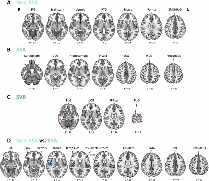
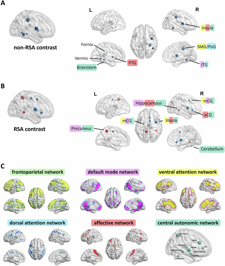
Major depressive disorder (MDD) is a heterogeneous, systemic disease often associated with cardiac autonomic dysfunction, yet neurophysiological mechanisms linking altered autonomic regulation to brain function remain largely unexplored to date. Here, we investigate functional brain correlates of cardiac autonomic modulation in MDD by integrating heart rate variability (HRV) with brain functional magnetic resonance imaging (fMRI). Forty participants (20 late-onset MDD patients, 20 healthy controls) undergo simultaneous electrocardiography and resting-state fMRI. HRV-derived autonomic regressors representing low-frequency and parasympathetic activity are estimated using time-varying bivariate autoregressive modeling and used to drive voxel-wise fMRI analysis via a general linear model. Group-level analyses assess diagnosis effects, controlling for age and sex. Post-hoc correlations are computed between HRV-related fMRI responses and clinical severity. Distinct patterns of brain-autonomic coupling emerge in MDD. Altered fMRI responses to autonomic dynamics are observed within central autonomic network regions, including the insula, cingulate gyrus, and hippocampus. The right insula shows consistent hypoactivation across multiple autonomic contrasts, with its response negatively correlating with depression severity. These findings provide preliminary evidence of altered brain-autonomic integration in MDD, particularly within interoceptive, self-referential, and affective networks. HRV-fMRI integration emerges as a promising multimodal framework for identifying multi-organ markers of cardiac autonomic dysfunction in psychiatric disorders.
Figures and tables
Reproduced without modification from: Goffi F, Enrico P, Reali P, Torino G, Marra MP, Di Consoli L, Ferro A, Schiena G, Torrente Y, Lombardi L, Triulzi FB, Bianchi AM, Brambilla P, Maggioni E (2026). Brain-heart interactions in late-onset major depressive disorder revealed by multimodal HRV-driven fMRI. Communications Biology. https://doi.org/10.1038/s42003-026-10156-5. Licensed under CC BY-NC-ND 4.0.